Preoperative Imaging in Predicting Post-Surgical Complications in Small Animals: Insights from In Vivo Studies
Article Sidebar
PDF downloads: 26
Crossref Citations: 0

Main Article Content
Abstract
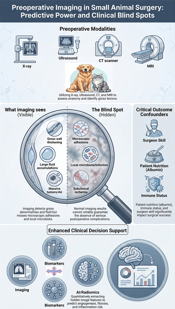
Preoperative imaging is a key tool in small-animal surgery; however, its predictive value for postoperative complications remains to be carefully investigated. The present study aimed to assess the predictive value and limitations of preoperative radiology for postoperative complications in small animals, based on evidence from in vivo studies. A systematic search of PubMed, Scopus, Web of Science, and Google Scholar databases was performed between 2020 and 2025 using keywords related to preoperative imaging, postoperative complications, dogs, cats, and animal models. Inclusion criteria included in vivo experimental studies on small animals (dogs and cats), reporting at least one preoperative imaging modality, including radiography, ultrasound, computed tomography (CT), and magnetic resonance imaging (MRI). Case reports, case series with a sample size of less than 5, and review articles were excluded. Out of 522 initial studies, duplicates were removed, and titles and abstracts were screened. This process resulted in 135 articles being fully evaluated, from which 116 studies were excluded, leaving 19 studies for inclusion. The positive predictive value of imaging for complications such as intestinal anastomotic leakage, pneumothorax after thoracotomy, and uroma after urinary tract surgery was moderate to acceptable. However, major limitations were present, including low sensitivity for detecting mild adhesions, inability to predict individual fibrotic responses, and failure to detect local microbiota. Additionally, normal preoperative radiological findings in small animals were associated with significant postoperative complications. Preoperative radiology in small animals has moderate to high predictive value for specific complications such as syringomyelia. However, owing to its limited sensitivity in detecting microscopic involvement and variations in individual tissue responses, preoperative radiology cannot serve as the only tool for clinical decision-making. Combining imaging with serum biomarkers and complementary laparoscopic evaluation are necessary for predicting post-surgical complications in small animals.
Article Details

This work is licensed under a Creative Commons Attribution 4.0 International License.
All claims expressed in this article are solely those of the authors and do not necessarily represent those of their affiliated organizations, or those of the publisher, the editors, and the reviewers. Any product that may be evaluated in this article, or claim that may be made by its manufacturer, is not guaranteed or endorsed by the publisher.
References
Ellison GW, Case JB, and Regier PJ. Intestinal surgery in small animals: Historical foundations, current thinking, and future horizons. Vet Surg. 2019; 48(7): 1171-1180. DOI: 10.1111/vsu.13275
Follette CM, Giuffrida MA, Balsa IM, Culp WT, Mayhew PD, Oblak ML, et al. A systematic review of criteria used to report complications in soft tissue and oncologic surgical clinical research studies in dogs and cats. Vet Surg. 2020; 49(1): 61-69. DOI: 10.1111/vsu.13279
Bojrab MJ, Waldron DR, and Toombs JP. Current techniques in small animal surgery, 5th ed. CRC Press; 2014. DOI: 10.1201/b17702
Nelson LL. Surgical site infections in small animal surgery. Vet Clin North Am Small Anim. Pract. 2011; 41(5): 1041-1056. DOI: 10.1016/j.cvsm.2011.05.010
Griffon D, and Hamaide A. Complications in small animal surgery. John Wiley & Sons, 2016. p. 8-14. DOI: 10.1002/9781119421344
Verwilghen D, and Singh A. Fighting surgical site infections in small animals: Are we getting anywhere?. Vet Clin North Am Small Anim Pract. 2015; 45(2): 243-276. DOI: 10.1016/j.cvsm.2014.11.001
Pailler S, Dolan ED, Slater MR, Gayle JM, Lesnikowski SM, DeClementi C, et al. Owner-reported long-term outcomes, quality of life, and longevity after hospital discharge following surgical treatment of pyometra in bitches and queens. J Am Vet Med Assoc. 2022; 260(2): 57-63. DOI: 10.2460/javma.20.12.0714
Greenhalgh SN, Reeve JA, Johnstone T, Goodfellow MR, Dunning MD, O'Neill EJ, et al. Long-term survival and quality of life in dogs with clinical signs associated with a congenital portosystemic shunt after surgical or medical treatment. J Am Vet Med Assoc. 2014; 245(5): 527-533. DOI: 10.2460/javma.245.5.527
Kipperman BS, Kass PH, and Rishniw M. Factors that influence small animal veterinarians' opinions and actions regarding cost of care and effects of economic limitations on patient care and outcome and professional career satisfaction and burnout. J Am Vet Med Assoc. 2017; 250(7): 785-794. DOI: 10.2460/javma.250.7.785
Moran CM, and Thomson AJ. Preclinical ultrasound imaging-a review of techniques and imaging applications. Front Phys. 2020; 8: 124. DOI: 10.3389/fphy.2020.00124
Kagadis GC, Loudos G, Katsanos K, Langer SG, and Nikiforidis GC. In vivo small animal imaging: Current status and future prospects. Med Phys. 2010; 37(12): 6421-6442. DOI: 10.1118/1.3515456
Yitbarek D, and Dagnaw GG. Application of advanced imaging modalities in veterinary medicine: A review. Vet Med Res Rep. 2022; 13: 117-130. DOI: 10.2147/VMRR.S367040
Gielen I, Caelenberg A, and Bree H. Clinical applications of computed tomography (CT) and magnetic resonance imaging (MRI) in small animals. Eur J Companion Anim Pract. 2012; 22(4): 84-103. Available at: http://hdl.handle.net/1854/LU-3091268
Ufuk F, Ocak İ, Chelala L, and Landeras L. Postoperative pulmonary complications: Clinical and imaging insights. Balkan Med J. 2025; 42(5): 405. DOI: 10.4274/balkanmedj.galenos.2025.2025-7.135
Safai Zadeh E, Görg C, Prosch H, Horn R, Jenssen C, and Dietrich CF. The role of thoracic ultrasound for diagnosis of diseases of the chest wall, the mediastinum, and the diaphragm-narrative review and pictorial essay. Diagnostics. 2023; 13(4): 767. DOI: 10.3390/diagnostics13040767
Vervoorn MT, Wulfse M, Mohamed Hoesein FA, Stellingwerf M, van der Kaaij NP, and de Heer LM. Application of three-dimensional computed tomography imaging and reconstructive techniques in lung surgery: A mini-review. Front Surg. 2022; 9: 1079857. DOI: 10.3389/fsurg.2022.1079857
Johnston SA, and Tobias KM. Veterinary surgery: Small animal. 2nd ed. Elsevier, 2017.
Mayhew PD. Complications of minimally invasive surgery in companion animals. Vet Clin North Am Small Anim Pract. 2011; 41(5): 1007-1021. DOI: 10.1016/j.cvsm.2011.05.008
Stieger‐Vanegas SM. Gastrointestinal imaging. Clinical small animal internal medicine. Wiley, 2020. p. 467-505. DOI: 10.1002/9781119501237.ch48
Kneissl SM, Prüllage ML, Vali Y, Vodnarek J, Klang A, Dolezal M, et al. Comparison of pre-and intraoperative findings in 35 cats and 60 dogs presenting with gastrointestinal signs. Front Vet Sci. 2025; 12: 1562792. DOI: 10.3389/fvets.2025.1562792
Boston S, and Henderson RA. Role of surgery in multimodal cancer therapy for small animals. Vet Clin North Am Small Anim Pract. 2014; 44(5): 855-870. DOI: 10.1016/j.cvsm.2014.05.008
Frey TN, Hoelzler MG, Scavelli TD, Fulcher RP, and Bastian RP. Risk factors for surgical site infection-inflammation in dogs undergoing surgery for rupture of the cranial cruciate ligament: 902 cases (2005-2006). J Am Vet Med Assoc. 2010; 236(1): 88-94. DOI: 10.2460/javma.236.1.88
Blondel M, Sonet J, Cachon T, Ségard‐Weisse E, Ferrand FX, and Carozzo C. Comparison of imaging techniques to detect migrating foreign bodies. Relevance of preoperative and intraoperative ultrasonography for diagnosis and surgical removal. Vet Surg. 2021; 50(4): 833-842. DOI: 10.1111/vsu.13607
Guiot LP, and Déjardin LM. Perioperative imaging in minimally invasive osteosynthesis in small animals. Vet Clin North Am Small Anim Pract. 2012; 42(5): 897-911. DOI: 10.1016/j.cvsm.2012.06.003
Moher D, Liberati A, Tetzlaff J, Altman DG, and Group P. Preferred reporting items for systematic reviews and meta-analyses: The PRISMA statement. Int J Surg. 2010; 8(5): 336-341. DOI: 10.1016/j.ijsu.2010.02.007
Alam IS, Steinberg I, Vermesh O, van den Berg NS, Rosenthal EL, van Dam GM, et al. Emerging intraoperative imaging modalities to improve surgical precision. Mol Imaging Biol. 2018; 20(5): 705-715. DOI: 10.1007/s11307-018-1227-6
Knight SR, Shaw CA, Pius R, Drake TM, Norman L, Ademuyiwa AO, et al. Global variation in postoperative mortality and complications after cancer surgery: A multicentre, prospective cohort study in 82 countries. Lancet. 2021; 397(10272): 387-397. DOI: 10.1016/S0140-6736(21)00001-5
Gans SL, Stoker J, and Boermeester MA. Plain abdominal radiography in acute abdominal pain; past, present, and future. Int J Gen Med. 2012; 5: 525-533. DOI: 10.2147/IJGM.S17410
von Stade L, and Sadar MJ. Advanced imaging of small mammals. Adv Small Anim Care. 2024; 5(1): 51-65. DOI: 10.1016/j.yasa.2024.06.004
Hölscher AH, Vallböhmer D, and Brabender J. The prevention and management of perioperative complications. Best Pract Res Clin Gastroenterol. 2006; 20(5): 907-923. DOI: 10.1016/j.bpg.2006.05.002
Tweedle E. Postoperative complications. In: Watson C, and Davies J, editors. Ellis and Calne's Lecture notes in general surgery. Wiley, 2023. p. 27-48. DOI: 10.1002/9781394322008.ch5
Cunha L, Horvath I, Ferreira S, Lemos J, Costa P, Vieira D, et al. Preclinical imaging: An essential ally in modern biosciences. Mol Diagn Ther. 2014; 18(2): 153-173. DOI: 10.1007/s40291-013-0062-3
Hurtig MB, Buschmann MD, Fortier LA, Hoemann CD, Hunziker EB, Jurvelin JS, et al. Preclinical studies for cartilage repair: Recommendations from the international cartilage repair society. Cartilage. 2011; 2(2): 137-152. DOI: 10.1177/1947603511401905
Hildebrandt IJ, Su H, and Weber WA. Anesthesia and other considerations for in vivo imaging of small animals. ILAR J. 2008; 49(1): 17-26. DOI: 10.1093/ilar.49.1.17
Van der Linden A, Van Camp N, Ramos‐Cabrer P, and Hoehn M. Current status of functional MRI on small animals: Application to physiology, pathophysiology, and cognition. NMR Biomed. 2007; 20(5): 522-545. DOI: 10.1002/nbm.1131
Albahrawy M, Abass M, Mosbah E, Karrouf G, and Zaghloul A. A narrative review on pathophysiology and the trends in preventing colon anastomotic leakage in animals. Mansoura Vet Med J. 2024; 25(2): 3. DOI: 10.35943/2682-2512.1235
Guyton KL, Hyman NH, and Alverdy JC. Prevention of perioperative anastomotic healing complications: Anastomotic stricture and anastomotic leak. Adv Surg. 2016; 50(1): 129. DOI: 10.1016/j.yasu.2016.03.011
Johnson C, Lapsley J, Piegols H, and Selmic L. Surgical approach and presence of preoperative pleural effusion impact thoracostomy tube usage in dogs and cats following thoracic surgery for suspected neoplasia. J Am Vet Med Assoc. 2024; 262(7): 1-7. DOI: 10.2460/javma.24.01.0005
Pawloski DR, and Broaddus KD. Pneumothorax: A review. J Am Anim Hosp Assoc. 2010; 46(6): 385-397. DOI: 10.5326/0460385
Epstein SE. Exudative pleural diseases in small animals. Vet Clin North Am Small Anim Pract. 2014; 44(1): 161-180. DOI: 10.1016/j.cvsm.2013.08.005
Lynch KC, Oliveira CR, Matheson JS, Mitchell MA, and O'Brien RT. Detection of pneumothorax and pleural effusion with horizontal beam radiography. Vet Radiol Ultrasound. 2012; 53(1): 38-43. DOI: 10.1111/j.1740-8261.2011.01854.x
Dickson R, Scharf VF, Michael AE, Walker M, Thomson C, Grimes J, et al. Surgical management and outcome of dogs with primary spontaneous pneumothorax: 110 cases (2009-2019). J Am Vet Med Assoc. 2021; 258(11): 1229-1235. DOI: 10.2460/javma.258.11.1229
Sériot P, Dunié‐Mérigot A, Tréhiou CB, Blond L, Bernardin F, Poujol L, et al. Treatment and outcome of spontaneous pneumothorax secondary to suspected migrating vegetal foreign body in 37 dogs. Vet Rec. 2021; 189(4): 22-41. DOI: 10.1002/vetr.22
Lamb CR. Veterinary diagnostic imaging: Probability, accuracy and impact. Vet J. 2016; 215: 55-63. DOI: 10.1016/j.tvjl.2016.03.017
Lamb CR, and David FH. Advanced imaging: Use and misuse. J Feline Med Surg. 2012; 14(7): 483-497. DOI: 10.1177/1098612X12451550
Chang JM, Lee HJ, Goo JM, Lee H-Y, Lee JJ, Chung J-K, et al. False positive and false negative FDG-PET scans in various thoracic diseases. Korean J Radiol. 2006; 7(1): 57-69. DOI: 10.3348/kjr.2006.7.1.57
Lauber DT, Fülöp A, Kovács T, Szigeti K, Máthé D, and Szijártó A. State of the art in vivo imaging techniques for laboratory animals. Lab Anim. 2017; 51(5): 465-478. DOI: 10.1177/0023677217695852
Balaban RS, and Hampshire VA. Challenges in small animal noninvasive imaging. ILAR J. 2001; 42(3): 248-262. DOI: 10.1093/ilar.42.3.248
Gabrielson K, Maronpot R, Monette S, Mlynarczyk C, Ramot Y, Nyska A, et al. In vivo imaging with confirmation by histopathology for increased rigor and reproducibility in translational research: A review of examples, options, and resources. ILAR J. 2018; 59(1): 80-98. DOI: 10.1093/ilar/ily010
de Brito Galvao JF, and Chew DJ. Metabolic complications of endocrine surgery in companion animals. Vet Clin North Am Small Anim Pract. 2011; 41(5): 847-868. DOI: 10.1016/j.cvsm.2011.05.012
Jones MA, MacCuaig WM, Frickenstein AN, Camalan S, Gurcan MN, Holter-Chakrabarty J, et al. Molecular imaging of inflammatory disease. Biomedicines. 2021; 9(2): 152. DOI: 10.3390/biomedicines9020152
Peñate Medina T, Kolb JP, Hüttmann G, Huber R, Peñate Medina O, Ha L, et al. Imaging inflammation-from whole body imaging to cellular resolution. Front Immunol. 2021; 12: 692222. DOI: 10.3389/fimmu.2021.692222
Koo V, Hamilton P, and Williamson K. Non‐invasive in vivo imaging in small animal research. Anal Cell Pathol. 2006; 28(4): 127-139. DOI: 10.1155/2006/245619
Bartling SH, Stiller W, Semmler W, and Kiessling F. Small animal computed tomography imaging. Curr Med Imaging. 2007; 3(1): 45-59. DOI: 10.2174/157340507779940327
Li H, Zhang H, Tang Z, and Hu G. Micro-computed tomography for small animal imaging: Technological details. Prog Nat Sci. 2008; 18(5): 513-521. DOI: 10.1016/j.pnsc.2008.01.002
Morrison ID, McLaughlin P, and Maher MM. Current status of imaging of the gastrointestinal tract: Imaging techniques and radiation issues. In: Adam A, Dixon AK, Gillard JH, editors. Grainger & Allison's diagnostic radiology: Abdominal imaging. 6th ed. Elsevier, 2015. p. 1-20.
Gerner-Rasmussen J, Donatsky AM, and Bjerrum F. The role of non-invasive imaging techniques in detecting intra-abdominal adhesions: A systematic review. Langenbecks Arch Surg. 2019; 404(6): 653-661. DOI: 10.1007/s00423-018-1732-8
Abdellatif A, Kramer M, Failing K, and Von Pückler K. Correlation between preoperative ultrasonographic findings and clinical, intraoperative, cytopathological, and histopathological diagnosis of acute abdomen syndrome in 50 dogs and cats. Vet Sci. 2017; 4(3): 39. DOI: 10.3390/vetsci4030039
Sturm MC, Abazid A, and Stope MB. Tissue adhesion after surgical interventions. Exp Ther Med. 2025; 29(5): 1-9. DOI: 10.3892/etm.2025.12847
Davey AK, and Maher PJ. Surgical adhesions: A timely update, a great challenge for the future. J Minim Invasive Gynecol. 2007; 14(1): 15-22. DOI: 10.1016/j.jmig.2006.07.013
Ellison GW. Complications of gastrointestinal surgery in companion animals. Vet Clin North Am Small Anim Pract. 2011; 41(5): 915-934. DOI: 10.1016/j.cvsm.2011.05.006
Holzman G, and Kleist TR, editors. Surgical patient care for veterinary technicians and nurses. 2nd ed. John Wiley & Sons, 2023. DOI: 10.1002/9781119760313
Antar SA, Ashour NA, Marawan ME, and Al-Karmalawy AA. Fibrosis: Types, effects, markers, mechanisms for disease progression, and its relation with oxidative stress, immunity, and inflammation. Int J Mol Sci. 2023; 24(4): 4004. DOI: 10.3390/ijms24044004
Jin Z, Guan Y, Yu G, and Sun Y. Magnetic resonance imaging of postoperative fracture healing process without metal artifact: A preliminary report of a novel animal model. Biomed Res Int. 2016; 2016(1): 1429892. DOI: 10.1155/2016/1429892
Low D, Treharne R, and Rutherford S. Machine‐learning prediction of postoperative complications after high tibial osteotomy for canine cranial cruciate ligament disease. Vet Surg. 2025; 54(7): 1286-1297. DOI: 10.1111/vsu.70007
Xue B, Li D, Lu C, King CR, Wildes T, and Avidan MS. Use of machine learning to develop and evaluate models using preoperative and intraoperative data to identify risks of postoperative complications. JAMA Net Open. 2021; 4(3): e212240. DOI: 10.1001/jamanetworkopen.2021.2240
Bouhali O, Bensmail H, Sheharyar A, David F, and Johnson JP. A review of radiomics and artificial intelligence and their application in veterinary diagnostic imaging. Vet Sci. 2022; 9(11): 620. DOI: 10.3390/vetsci9110620
Tomaszewski MR, and Gillies RJ. The biological meaning of radiomic features. Radiology. 2021; 298(3): 505-516. DOI: 10.1148/radiol.2021202553
Rajgor A, Gill T, Aboagye E, Mill A, Rushton S, Obara B, et al. Radiomics from routine CT and PET/CT imaging in laryngeal squamous cell carcinoma: A systematic review with radiomics quality score assessment. Cancers. 2026; 18(2): 237. DOI: 10.3390/cancers18020237
Bonde A, Varadarajan KM, Bonde N, Troelsen A, Muratoglu OK, Malchau H, et al. Assessing the utility of deep neural networks in predicting postoperative surgical complications: A retrospective study. Lancet Digit Health. 2021; 3(8): 471-485. DOI: 10.1016/S2589-7500(21)00084-4
Hussain SM, Brunetti A, Lucarelli G, Memeo R, Bevilacqua V, and Buongiorno D. Deep learning based image processing for robot assisted surgery: A systematic literature survey. IEEE Access. 2022; 10: 122627-122657. DOI: 10.1109/ACCESS.2022.3223704
Scholz AM, Bünger L, Kongsro J, Baulain U, and Mitchell AD. Non-invasive methods for the determination of body and carcass composition in livestock: Dual-energy X-ray absorptiometry, computed tomography, magnetic resonance imaging and ultrasound: Invited review. Animal. 2015; 9(7): 1250-1264. DOI: 10.1017/S1751731115000336
Song Q, He X, Wang Y, Gao H, Tan L, and Ma J. Clinical validation of AI assisted animal ultrasound models for diagnosis of early liver trauma. Sci Rep. 2025; 15(1): 22513. DOI: 10.1038/s41598-025-91900-5
Leivaditis V, Maniatopoulos AA, Lausberg H, Mulita F, Papatriantafyllou A, Liolis E, et al. Artificial intelligence in thoracic surgery: A review bridging innovation and clinical practice for the next generation of surgical care. J Clin Med. 2025; 14(8): 2729. DOI: 10.3390/jcm14082729